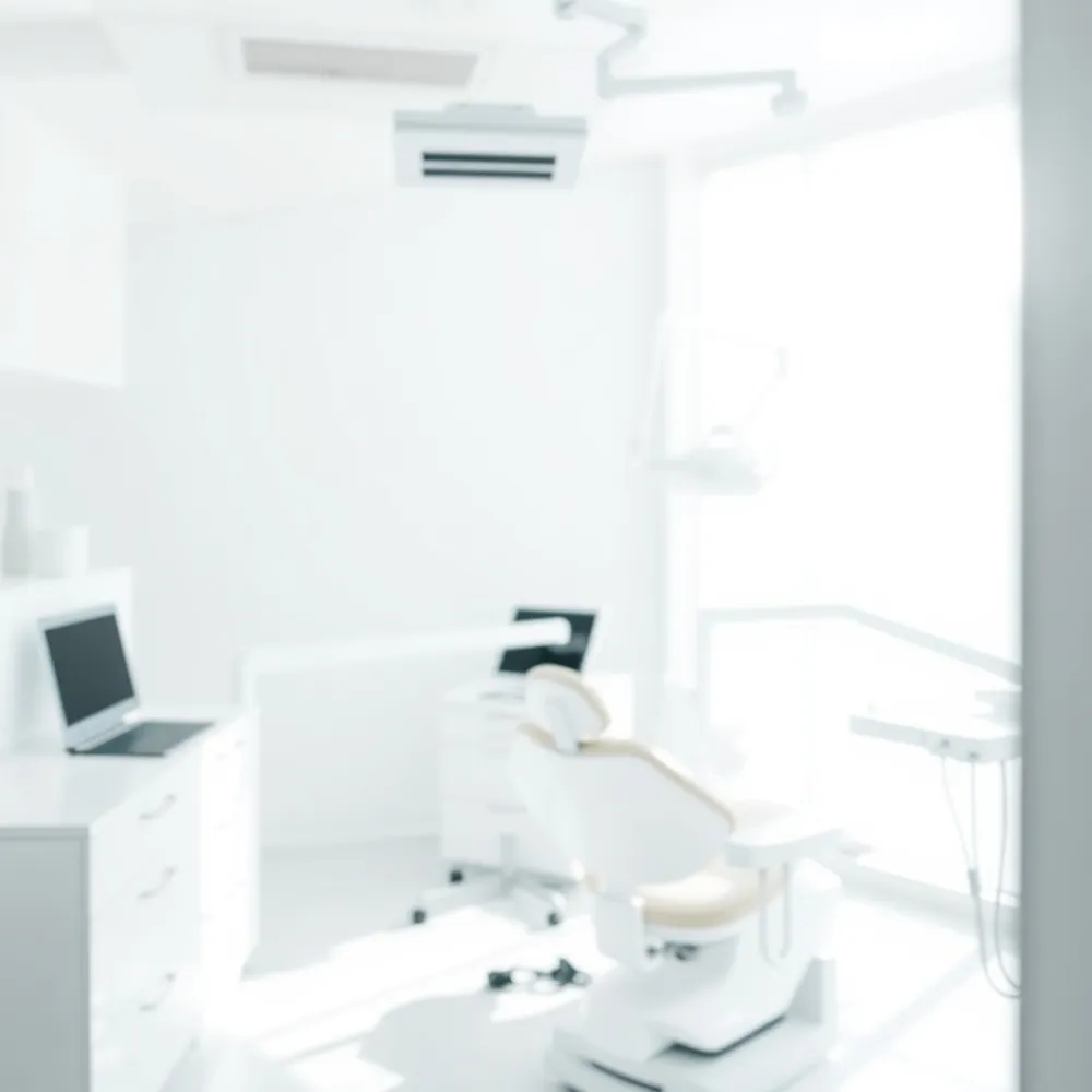
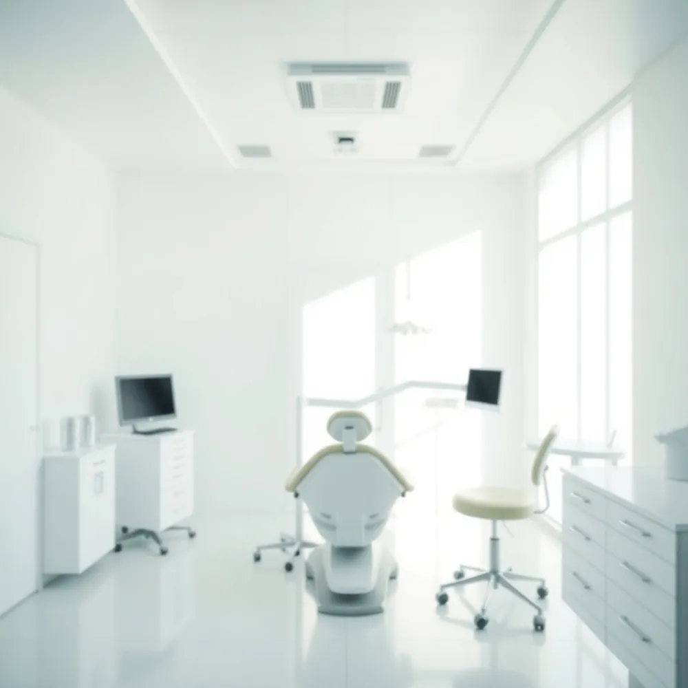
A New Era in Dental Imaging
Digital X‑ray technology replaces film with electronic sensors that capture high‑resolution images in seconds. The sensors use far less ionizing radiation—often 60‑90 % lower than traditional film—making scans safer for children, frequent visitors, and pregnant patients. Immediate image processing lets the dentist view, enhance (contrast, brightness, zoom) and discuss findings with the patient during the same appointment, reducing chair‑time and the need for repeat exposures. Our practice embraces this cutting‑edge, patient‑centered approach by integrating digital radiographs with electronic health records, enabling seamless sharing with specialists and secure, long‑term storage. By offering faster, safer diagnostics, we meet modern expectations for transparent communication, early detection of decay or bone loss, and personalized treatment planning—all while minimizing radiation, waste, and environmental impact. This commitment to advanced imaging ensures families receive the highest standard of preventive and restorative dental care.
Radiation Safety and the 3‑3‑3 Rule
![]() Digital sensors cut radiation by 80‑90 % versus film, so a single intra‑oral bite‑wing or periapical image delivers only 0.1–0.8 mrem—far less than a day of natural background exposure. This tiny dose makes routine lead aprons unnecessary; modern guidelines (AAOS, ACR) deem them redundant because the beam is tightly collimated and the ALARA principle is met. Faster‑speed film historically reduced exposure by 30‑60 %, but digital radiography now achieves even greater safety without sacrificing image quality. The smallest dose comes from digital intra‑oral X‑rays; panoramic or CBCT scans emit much more. For everyday oral hygiene, the 3‑3‑3 rule reminds patients to brush three times a day for three minutes and wait three hours after meals before bedtime, giving saliva a chance to protect enamel. In short, digital X‑rays are safer, faster, and more comfortable, supporting preventive care for families.
Digital sensors cut radiation by 80‑90 % versus film, so a single intra‑oral bite‑wing or periapical image delivers only 0.1–0.8 mrem—far less than a day of natural background exposure. This tiny dose makes routine lead aprons unnecessary; modern guidelines (AAOS, ACR) deem them redundant because the beam is tightly collimated and the ALARA principle is met. Faster‑speed film historically reduced exposure by 30‑60 %, but digital radiography now achieves even greater safety without sacrificing image quality. The smallest dose comes from digital intra‑oral X‑rays; panoramic or CBCT scans emit much more. For everyday oral hygiene, the 3‑3‑3 rule reminds patients to brush three times a day for three minutes and wait three hours after meals before bedtime, giving saliva a chance to protect enamel. In short, digital X‑rays are safer, faster, and more comfortable, supporting preventive care for families.
Instant Diagnosis and Real‑Time Patient Communication
![]() What are the advantages of digital imaging in dentistry? Digital imaging delivers instant, high‑resolution images with low radiation, eliminating film processing, reducing waste, and enabling easy enhancement and sharing for better communication and confidence.
What are the advantages of digital imaging in dentistry? Digital imaging delivers instant, high‑resolution images with low radiation, eliminating film processing, reducing waste, and enabling easy enhancement and sharing for better communication and confidence.
What are three advantages of digital X‑rays over film X‑rays? Sharper, zoomable images; up to 90 % lower radiation dose; immediate electronic access that speeds diagnosis and reduces repeat exposures.
Are dental X‑rays safer now than in the past? Yes—modern sensors, collimation and the ALARA principle have cut doses to levels comparable to a few minutes of natural background radiation, making them a safe, routine diagnostic tool.
Environmental and Practice‑Efficiency Benefits
![]() Digital dental X‑rays eliminate the need for chemical processing, so the toxic developers and fixers that once created hazardous waste are no longer used. This dramatically reduces a practice’s environmental footprint and removes the health risks associated with chemical handling. Because no film is required, costs for purchasing, processing, and storing physical radiographs disappear, freeing up office space and cutting supply expenses. The instant electronic capture and display of images streamlinelines the workflow: clinicians can review scans within seconds, adjust contrast or zoom on the screen, and discuss findings with patients during the same visit, which shortens chair‑time and improves patient comfort. All images are archived electronically, allowing secure backup, rapid retrieval, and easy sharing with specialists. Integration with practice‑management software and electronic health‑record (EHR) systems further automates documentation, reduces paperwork, and supports seamless coordination of care—all while supporting a greener, more cost‑effective dental practice.
Digital dental X‑rays eliminate the need for chemical processing, so the toxic developers and fixers that once created hazardous waste are no longer used. This dramatically reduces a practice’s environmental footprint and removes the health risks associated with chemical handling. Because no film is required, costs for purchasing, processing, and storing physical radiographs disappear, freeing up office space and cutting supply expenses. The instant electronic capture and display of images streamlinelines the workflow: clinicians can review scans within seconds, adjust contrast or zoom on the screen, and discuss findings with patients during the same visit, which shortens chair‑time and improves patient comfort. All images are archived electronically, allowing secure backup, rapid retrieval, and easy sharing with specialists. Integration with practice‑management software and electronic health‑record (EHR) systems further automates documentation, reduces paperwork, and supports seamless coordination of care—all while supporting a greener, more cost‑effective dental practice.
Advanced Clinical Applications and AI Assistance
![]() Digital X‑rays now serve as a gateway to cutting‑edge dental care. By linking the 2‑D sensor to 3‑D cone‑beam CT, clinicians can overlay the radiograph on a volumetric scan, enabling precise implant planning and accurate assessment of bone anatomy before surgery. AI‑driven algorithms automatically highlight early caries, subtle bone loss, and periapical lesions, giving the dentist a second set of eyes that speeds diagnosis and reduces missed pathology. Secure, encrypted transmission lets these enhanced images travel instantly to remote specialists, supporting tele‑dentistry consultations without compromising patient privacy. When a restoration is needed, the same digital file feeds directly into computer‑aided design (CAD) software, allowing same‑day milling of crowns, veneers or inlays and minimizing visits. Looking ahead, predictive analytics will mine longitudinal radiographic data to forecast disease progression, helping families adopt preventive measures before problems arise. Together, these innovations make dental treatment safer, faster, and more personalized.
Digital X‑rays now serve as a gateway to cutting‑edge dental care. By linking the 2‑D sensor to 3‑D cone‑beam CT, clinicians can overlay the radiograph on a volumetric scan, enabling precise implant planning and accurate assessment of bone anatomy before surgery. AI‑driven algorithms automatically highlight early caries, subtle bone loss, and periapical lesions, giving the dentist a second set of eyes that speeds diagnosis and reduces missed pathology. Secure, encrypted transmission lets these enhanced images travel instantly to remote specialists, supporting tele‑dentistry consultations without compromising patient privacy. When a restoration is needed, the same digital file feeds directly into computer‑aided design (CAD) software, allowing same‑day milling of crowns, veneers or inlays and minimizing visits. Looking ahead, predictive analytics will mine longitudinal radiographic data to forecast disease progression, helping families adopt preventive measures before problems arise. Together, these innovations make dental treatment safer, faster, and more personalized.
Patient‑Friendly Oral‑Health Routines
![]() What is the 2‑2‑2 rule for teeth?
The 2‑2‑2 rule is a simple, evidence‑based guideline for maintaining healthy teeth and gums. It recommends brushing your teeth twice each day, spending at least two minutes on each brushing session to effectively remove plaque. In addition, you should schedule a professional dental exam and cleaning twice a year so that a dentist can catch problems early and remove tartar that home care cannot. Following these three steps helps prevent cavities, gum disease, and bad breath while supporting overall oral health.
What is the 2‑2‑2 rule for teeth?
The 2‑2‑2 rule is a simple, evidence‑based guideline for maintaining healthy teeth and gums. It recommends brushing your teeth twice each day, spending at least two minutes on each brushing session to effectively remove plaque. In addition, you should schedule a professional dental exam and cleaning twice a year so that a dentist can catch problems early and remove tartar that home care cannot. Following these three steps helps prevent cavities, gum disease, and bad breath while supporting overall oral health.
Comfort advantages of small, wireless digital sensors Modern digital X‑ray sensors are tiny, lightweight, and often wireless, making them far more comfortable than bulky film plates. Their slim profile reduces gag reflex and allows quicker placement, improving the patient experience during routine imaging.
Child and pregnant‑patient safety with low‑dose imaging Digital X‑rays emit 60‑90 % less radiation than traditional film, keeping exposure well below natural background levels. This low dose is especially important for children and pregnant patients, aligning with the ALARA principle and often eliminating the need for lead aprons.
Aesthetic smile design and the 50‑40‑30 rule The 50‑40‑30 rule describes the ideal proportion of visible tooth widths in a harmonious smile: central incisors occupy about 50 % of the incisal display, central‑to‑lateral incisor width about 40 %, and lateral‑to‑canine width about 30 %. Digital imaging lets clinicians plan and preview these proportions for personalized, beautiful results.
Embracing Safer, Faster, and Smarter Dental Care
Digital X‑rays give families eight surprising benefits: up to 90 % lower radiation, instant images that shorten visits, high‑resolution pictures that spot tiny cavities, adjustable contrast and zoom for precise diagnosis, electronic storage that prevents loss and eases sharing, elimination of chemical waste for a greener office, reusable sensors that boost comfort and reduce costs, and seamless integration with electronic health records for coordinated care. These advantages echo Dr. Parrella’s patient‑first philosophy by prioritizing safety, transparency and efficiency. We invite Somerville families to experience our modern, low‑dose imaging—quick, comfortable, and environmentally responsible—so every visit supports lasting oral health. Schedule your appointment today and see the difference firsthand.