Welcome to Your First Digital X‑Ray Visit
Digital dental X‑rays are now the standard of care because they use up to 90 % less radiation than traditional film, delivering high‑resolution images while adhering to the ALARA principle for patient safety. The appointment is quick and painless—after you remove jewelry or metal objects, a lead apron (and, when needed, a thyroid collar) is placed for extra protection. A small, smooth sensor is gently positioned in your mouth; you may be asked to bite down briefly and stay still for a few seconds while the image is captured. The entire process, from preparation to image review, typically takes less than ten minutes, and you can resume normal activities immediately. Our team monitors your comfort, explains each step, and uses instant digital images so the dentist can discuss findings with you on the spot, ensuring a clear, compassionate, and safe experience.
Preparing for Your Appointment
![]() Before your digital dental X‑ray, start by removing any metal—jewelry, glasses, dentures, or hair accessories—so they don’t interfere with image quality. A quick brush of your teeth isn’t required, but a clean mouth helps produce clearer pictures and keeps the sensor area sanitary. Most offices, including Dr. Anthony P. Parrella’s practice at 102 College Avenue, Somerville, MA 02144, will provide a lead apron (and, if needed, a thyroid collar) to protect you from scatter radiation; the dose from a digital X‑ray is only a fraction of everyday background radiation.
Before your digital dental X‑ray, start by removing any metal—jewelry, glasses, dentures, or hair accessories—so they don’t interfere with image quality. A quick brush of your teeth isn’t required, but a clean mouth helps produce clearer pictures and keeps the sensor area sanitary. Most offices, including Dr. Anthony P. Parrella’s practice at 102 College Avenue, Somerville, MA 02144, will provide a lead apron (and, if needed, a thyroid collar) to protect you from scatter radiation; the dose from a digital X‑ray is only a fraction of everyday background radiation.
Do I need to brush my teeth before a dental X‑ray? A gentle brush can improve image clarity, though it isn’t mandatory.
Can you request dental X‑rays from your dentist? Yes—under HIPAA you may obtain copies at no charge, either digitally or on film.
How many dental X‑rays are safe in a month? Modern digital sensors use ultra‑low radiation; a few clinically justified X‑rays in a month are safe, especially when lead aprons and proper collimation are used.
Radiation Dose, Safety Principles, and Coverage
![]() Digital dental X‑rays expose patients to only a tiny fraction of the radiation we encounter daily. A single bite‑wing is about 0.02 mSv (≈20 µSv), comparable to a few days of natural background radiation; a periapical image adds roughly 0.005 mSv, and a full‑mouth series of four bite‑wings totals about 0.02 mSv. Panoramic scans range from 0.0047 mSv to 0.0143 mSv, while a digital full‑mouth series is about 0.09 mSv. The ALARA (As Low As Reasonably Achievable) principle guides every exposure, keeping dose as low as possible while still providing diagnostic quality. The primary risks are a very small increase in lifetime cancer probability, especially head‑and‑neck or thyroid cancers, and cumulative exposure from repeated exams—particularly in children or pregnant patients. Modern digital sensors, lead aprons, and thyroid collars markedly reduce these hazards. Insurance wise, MassHealth covers dental crowns when medically necessary, and most private plans cover preventive X‑rays at 100 % when justified. The informal “2‑year rule” limits the interval between dental visits for insurance eligibility, but clinicians usually recommend cleanings every six months or more often for high‑risk patients.
Digital dental X‑rays expose patients to only a tiny fraction of the radiation we encounter daily. A single bite‑wing is about 0.02 mSv (≈20 µSv), comparable to a few days of natural background radiation; a periapical image adds roughly 0.005 mSv, and a full‑mouth series of four bite‑wings totals about 0.02 mSv. Panoramic scans range from 0.0047 mSv to 0.0143 mSv, while a digital full‑mouth series is about 0.09 mSv. The ALARA (As Low As Reasonably Achievable) principle guides every exposure, keeping dose as low as possible while still providing diagnostic quality. The primary risks are a very small increase in lifetime cancer probability, especially head‑and‑neck or thyroid cancers, and cumulative exposure from repeated exams—particularly in children or pregnant patients. Modern digital sensors, lead aprons, and thyroid collars markedly reduce these hazards. Insurance wise, MassHealth covers dental crowns when medically necessary, and most private plans cover preventive X‑rays at 100 % when justified. The informal “2‑year rule” limits the interval between dental visits for insurance eligibility, but clinicians usually recommend cleanings every six months or more often for high‑risk patients.
Types of Digital Dental X‑Rays and Their Uses
![]() Digital dental radiography includes two main categories: intra‑oral imaging (sensor placed inside the mouth) and extra‑oral imaging (sensor outside the mouth, such as panoramic scans). Intra‑oral exams cover periapical X‑rays—full‑tooth images used to assess root health, infections, bone levels, and canal anatomy—and bitewing X‑rays, which capture upper and lower posterior teeth together to reveal interproximal cavities and bone loss. Extra‑oral options like panoramic and cone‑beam CT provide a broad view of the jaws and sinuses for implant planning or orthodontics. The first digital dental radiographic system, RadioVisioGraphy, debuted in the early 1990s, introducing solid‑state sensors and instant image transfer. Today, most dental offices—including family practices such as Dr. Parrella’s—use digital X‑rays benefiting from up to 80 % lower radiation, immediate high‑resolution images, and seamless electronic storage and sharing.
Digital dental radiography includes two main categories: intra‑oral imaging (sensor placed inside the mouth) and extra‑oral imaging (sensor outside the mouth, such as panoramic scans). Intra‑oral exams cover periapical X‑rays—full‑tooth images used to assess root health, infections, bone levels, and canal anatomy—and bitewing X‑rays, which capture upper and lower posterior teeth together to reveal interproximal cavities and bone loss. Extra‑oral options like panoramic and cone‑beam CT provide a broad view of the jaws and sinuses for implant planning or orthodontics. The first digital dental radiographic system, RadioVisioGraphy, debuted in the early 1990s, introducing solid‑state sensors and instant image transfer. Today, most dental offices—including family practices such as Dr. Parrella’s—use digital X‑rays benefiting from up to 80 % lower radiation, immediate high‑resolution images, and seamless electronic storage and sharing.
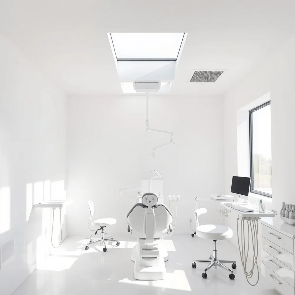
The In‑Office Digital X‑Ray Procedure
![]() During a digital dental X‑ray the patient removes jewelry, watches or metal objects that could blur the image. A lead apron or thyroid collar may be placed for protection, though sensors already keep the dose low. The technologist positions a small bite‑size sensor inside the mouth, using a bite block or mirror, while the patient gently bites down. For intraoral shots the patient holds still and briefly stops breathing for one second to avoid motion blur; extraoral panoramic views require the head to stay still while the arm rotates. The X‑ray beam fires for a fraction of a second and the sensor instantly sends a high‑resolution image to the computer, where the dentist reviews it with the patient in real time. Costs vary: a bitewing $25‑$50, a periapical $30‑$60, a panoramic $80‑$150, and a full‑mouth series $150‑$250. Insurance often covers preventive films, and many offices bundle the fee into the exam cost.
During a digital dental X‑ray the patient removes jewelry, watches or metal objects that could blur the image. A lead apron or thyroid collar may be placed for protection, though sensors already keep the dose low. The technologist positions a small bite‑size sensor inside the mouth, using a bite block or mirror, while the patient gently bites down. For intraoral shots the patient holds still and briefly stops breathing for one second to avoid motion blur; extraoral panoramic views require the head to stay still while the arm rotates. The X‑ray beam fires for a fraction of a second and the sensor instantly sends a high‑resolution image to the computer, where the dentist reviews it with the patient in real time. Costs vary: a bitewing $25‑$50, a periapical $30‑$60, a panoramic $80‑$150, and a full‑mouth series $150‑$250. Insurance often covers preventive films, and many offices bundle the fee into the exam cost.
Interpreting Results, Follow‑Up, and Ongoing Care
![]() After the sensor captures the image, a radiologist reads the digital radiograph and flags any abnormalities. The report is sent to the treating dentist, who combines it with clinical exam. AI algorithms can highlight cavities, bone loss or root issues, helping the dentist explain the picture. Most practices deliver the report within 24–48 hours; some show the image instantly on a monitor and discuss it during the same visit. Patients are told what the images show, why treatment may be needed, and steps. Follow‑up appointments are based on findings—often a six‑month recall, but the informal “2‑year rule” advises no more than two years between visits for adults. Two‑fold risks of dental radiography are a slight increase in lifetime cancer risk and cumulative exposure, especially for children or pregnant patients; shielding and low‑dose sensors keep these risks minimal. MassHealth covers crowns when medically necessary, so eligible members can receive restorative care without extra cost.
After the sensor captures the image, a radiologist reads the digital radiograph and flags any abnormalities. The report is sent to the treating dentist, who combines it with clinical exam. AI algorithms can highlight cavities, bone loss or root issues, helping the dentist explain the picture. Most practices deliver the report within 24–48 hours; some show the image instantly on a monitor and discuss it during the same visit. Patients are told what the images show, why treatment may be needed, and steps. Follow‑up appointments are based on findings—often a six‑month recall, but the informal “2‑year rule” advises no more than two years between visits for adults. Two‑fold risks of dental radiography are a slight increase in lifetime cancer risk and cumulative exposure, especially for children or pregnant patients; shielding and low‑dose sensors keep these risks minimal. MassHealth covers crowns when medically necessary, so eligible members can receive restorative care without extra cost.
Your Next Steps After the X‑Ray
After the digital X‑ray, the dentist will pull up the high‑resolution image on the screen and walk you through what they see—showing any cavities, bone loss, or other findings in real time. This visual review helps you understand the diagnosis and why a particular treatment is recommended, whether it’s a simple filling, a periodontal plan, or a referral for a specialist. Because the image is stored electronically, the dentist can compare it to previous radiographs and track changes over time. Before you leave, discuss the timeline for any needed procedures and set up follow‑up appointments, including routine check‑ups or a repeat X‑ray in 6–18 months, to keep your oral health on track.