Embracing Modern Technology for Your Family's Smile
For over a century, traditional film X-rays have helped dentists find cavities, monitor jaw growth, and spot infections. Digital X-rays now offer a much safer and more capable alternative. Our practice is committed to using the safest technology available, providing lower radiation, instant images, and more accurate diagnoses for your family.
What Are Digital Dental X-Rays?
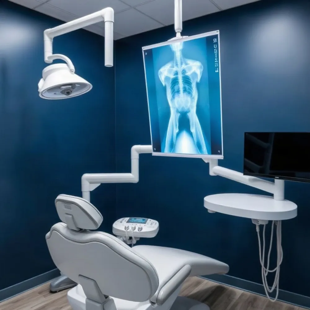 Digital radiography in dentistry uses electronic sensors rather than photographic film to capture detailed images of teeth, gums, and surrounding bone structures. When the sensor is placed in the mouth, it converts X-ray energy into an electrical signal that is transmitted directly to a computer, where the image appears on screen within seconds. This instant process eliminates the need for chemical film development and allows the dental team to diagnose issues more efficiently.
There are three methods of acquiring digital images: the direct method employs a solid-state sensor placed in the mouth; the indirect method uses a scanner to convert traditional X-ray film into a digital file; and the semi-indirect method combines a sensor and scanner. Most modern practices, including Dr. Parrella's, rely on direct digital sensors because they provide the fastest results and highest image quality while reducing radiation exposure by up to 90% compared to conventional film X-rays.
Intraoral and Extraoral Digital X-Rays
Digital X-rays fall into two main categories based on where the sensor is placed. Intraoral X-rays are taken inside the mouth and include bitewing X-rays (which detect cavities between teeth and below the gumline) and periapical X-rays (which capture the entire tooth from crown to root, helping identify abscesses, bone loss, and infections). These are the most common type taken during routine checkups.
Extraoral X-rays are taken outside the mouth and provide a broader view of the jaw and facial structures. They include panoramic X-rays (which show the entire mouth in a single image, useful for planning implants and detecting impacted wisdom teeth), cephalometric projections (used for orthodontic treatment planning), and cone beam CT scans (which create 3D images for evaluating bone for implants or identifying facial bone problems).
| X-Ray Type | Category | Primary Use |
|---|---|---|
| Bitewing | Intraoral | Detect cavities between teeth and below gumline |
| Periapical | Intraoral | View entire tooth, root, and surrounding bone |
| Occlusal | Intraoral | Reveal impacted teeth and jaw fractures |
| Panoramic | Extraoral | Single image of full mouth; implant and wisdom tooth planning |
| Cephalometric | Extraoral | Side view of head for orthodontic planning |
| Cone beam CT | Extraoral | 3D images for implant placement and bone evaluation |
Digital vs. Traditional: A Clear Advantage
When you visit the dentist, the type of X-ray technology used directly impacts your safety, comfort, and the quality of your diagnosis. Traditional film X-rays have served dentistry well for decades, but digital dental X-rays have become the clear standard for modern, patient-centered care. The differences are substantial, from radiation exposure to diagnostic capabilities.
The most significant advantage of digital X-rays is radiation safety. As noted by the FDA, lower radiation dose is a key benefit of newer digital systems over older film-based ones, following the ALARA (As Low As Reasonably Achievable) principle. Studies consistently show that digital X-rays can reduce radiation exposure by up to 80–90% compared to traditional film. In our practice at drparrella.com, we prioritize this safety benefit, especially for children and families who may require more frequent monitoring.
Time is another clear differentiator. A digital sensor captures an image and displays it on a screen within seconds, eliminating the minutes-long chemical processing required for film in a darkroom. This efficiency means shorter appointments and quicker diagnoses, allowing dentists to immediately assess image quality and avoid retakes. Traditional film cannot be reviewed until development is complete, sometimes necessitating a second appointment if the image is unusable.
Image Quality and Diagnostic Power
Digital X-rays. Provide superior, high-resolution images that can be instantly enhanced on a computer screen. Dentists can zoom in, adjust contrast and brightness, and apply color filters to detect early cavities, bone loss, or infections that might be missed on film. This manipulation leads to more accurate diagnoses and better treatment planning.
Traditional Film X-rays. Offer excellent picture quality with high resolution, and their images cannot be digitally altered, which some consider an advantage for legal records. However, the inability to enhance details can make it harder to spot subtle early-stage problems, and issues like over- or under-exposure require a completely new image.
For patients, the enhanced clarity of digital images also improves communication. As reported in a PMC study, the ability to show magnified, adjusted images on a chairside monitor helps patients understand their oral health conditions more clearly, building trust and enabling informed decisions about their treatment.
Environmental and Practical Benefits
Digital radiography is inherently more eco-friendly. It eliminates the need for photographic film, lead foil, and the toxic chemical developers and fixers required for processing film. This reduces hazardous waste in dental offices. At drparrella.com, this aligns with a commitment to a healthier environment for the Somerville community.
Digital records also solve the storage and sharing challenges of film. Physical film requires bulky filing cabinets and can be lost or damaged. Digital images are stored securely in electronic patient records, can be instantly retrieved for comparison with previous visits, and shared with specialists or insurance companies via encrypted email. If a patient needs to see a specialist, digital X-rays can be sent ahead, eliminating the need for duplicate exposures.
While traditional film systems have lower upfront equipment costs and some patients find the thinner film sensors slightly more comfortable, these advantages are outweighed by the significant radiation reduction, diagnostic enhancement, and workflow efficiency of digital systems. Modern digital sensors are increasingly designed for patient comfort, and at drparrella.com, our team is trained to ensure a gentle, efficient experience. For families seeking the safest and most accurate care, digital X-rays are the clear choice.
Understanding Safety and Radiation Levels
Patient safety is at the heart of modern dentistry, and the move to digital X-ray technology has made diagnostic imaging remarkably safe. Digital systems use electronic sensors instead of film, requiring up to 90% less radiation than traditional film-based X-rays, as noted in a study on digital radiography. This reduction directly aligns with the ALARA (As Low As Reasonably Achievable) principle, a core guideline set by the FDA for minimizing radiation exposure. At our practice, we follow this principle strictly, ensuring each X-ray is justified and optimized for the lowest possible dose. We also use protective lead aprons and thyroid collars for added security, especially for children.
To understand just how low the exposure is, consider the numbers. A single digital bitewing X-ray, the type commonly taken during checkups, delivers about 0.002 to 0.006 mSv. This is comparable to the natural background radiation you absorb in a few hours or the radiation from eating roughly 2 to 5 bananas. A full-mouth series of 18 digital X-rays totals about 0.023 mSv, which is equivalent to about 1 to 3 days of natural background radiation.
These levels are remarkably small when compared to other common medical imaging procedures. A standard chest X-ray is about 0.1 mSv, which is 20 times the radiation of a single dental digital X-ray. A CT scan of the abdomen and pelvis delivers about 10 mSv, or about 2,000 times the dose. For reference, the average person in the U.S. receives about 3 mSv per year from natural background radiation alone. This context shows that a routine dental X-ray is an extremely low-risk procedure, especially for children and families who prioritize safety.
The American Dental Association has updated its guidelines based on this data. As of 2024, lead aprons and thyroid collars are no longer recommended for routine dental X-rays because modern digital equipment uses such focused, low-dose beams that exposure to other body parts is negligible. At our Somerville office, we always follow the latest safety protocols and take X-rays only when they provide clear diagnostic value for your care. We will never recommend an X-ray without a specific reason, and we keep detailed records to avoid unnecessary repeats.
The Advantages of Going Digital
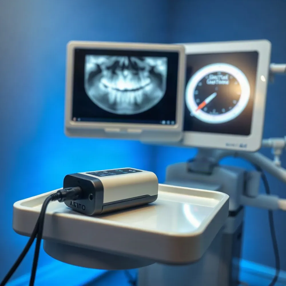 When digital X-rays replaced film in dental offices, the experience changed for both clinician and patient. The first and most dramatic improvement is speed: a sensor inside the mouth transmits the image to a computer screen in about five seconds, eliminating the minutes-long wait for film development. That faster turnaround means quicker diagnosis and less time in the chair.
Beyond speed, digital sensors capture images at a much higher resolution than film. The dentist can zoom in on a suspicious area, adjust brightness and contrast, or apply color filters to reveal details that might otherwise stay hidden. This makes it easier to spot early cavities, bone loss, infections, and root abnormalities before they become serious problems.
The same high-quality images also become a powerful educational tool. Patients can see on a monitor exactly what the dentist sees — a cavity forming between teeth, the extent of gum disease, or the position of an impacted tooth. Visual aids like these help build trust and turn a technical consultation into a shared conversation about oral health.
Environment. Digital radiography eliminates the chemical processing and hazardous waste associated with film, making it a more eco-friendly choice.
Storage and sharing. Images are stored electronically in patient records and can be transmitted securely to specialists or insurance companies. This reduces the need for repeat X-rays when visiting a new provider.
At Dr. Parrella's practice in Somerville, these advantages translate directly into a safer, faster, and more transparent experience for every patient. The instant, high-resolution images allow the team to identify issues early and explain them clearly, supporting the kind of compassionate, preventive care that families in the community have come to expect.
Putting Patient Safety First in Somerville
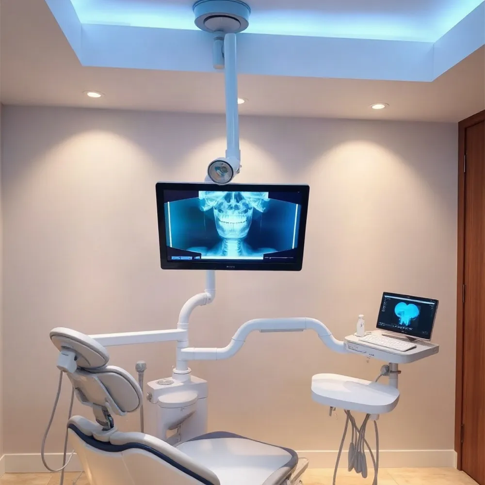 For families in Somerville, MA, choosing a dental provider means trusting that every precaution is taken. At our practice, we use state-of-the-art digital X-ray systems for every exam. This choice aligns with the ALARA (As Low As Reasonably Achievable) principle, a core radiation safety standard endorsed by the FDA that aims to minimize exposure while delivering the diagnostic information needed.
Our family-focused approach means we prioritize faster, more comfortable procedures. Because digital sensors capture images in seconds and eliminate the need for film development, chair time is shorter. This is especially valuable for children and patients with anxiety, as they spend less time holding still with a sensor in place.
We take X-rays only when there is a clear diagnostic need, and we never perform them as a routine. Digital records allow for seamless continuity of care with specialists. If a referral is required, we can email your images directly to the specialist’s office, avoiding the need for repeat exposures.
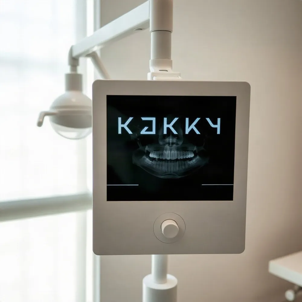
Patients are partners in their care. After each image is captured, we display it on a monitor beside the chair and explain what we see. This real-time discussion helps you understand your oral health without needing a dental degree, building trust and supporting confident treatment decisions.
A Brighter, Safer Future for Your Dental Health
Digital X-rays have become a cornerstone of modern, patient-centered dentistry. The combination of lower radiation and clearer images allows dentists to detect problems earlier than ever before, often catching decay or bone loss before symptoms appear.
At drparrella.com, we are committed to providing the highest standard of care with the safest technology available. We encourage every patient to ask questions about their X‑rays during their visit — understanding what the images show helps build trust and empowers you to take an active role in your oral health.