Embracing Advanced Dental Care for Your Family
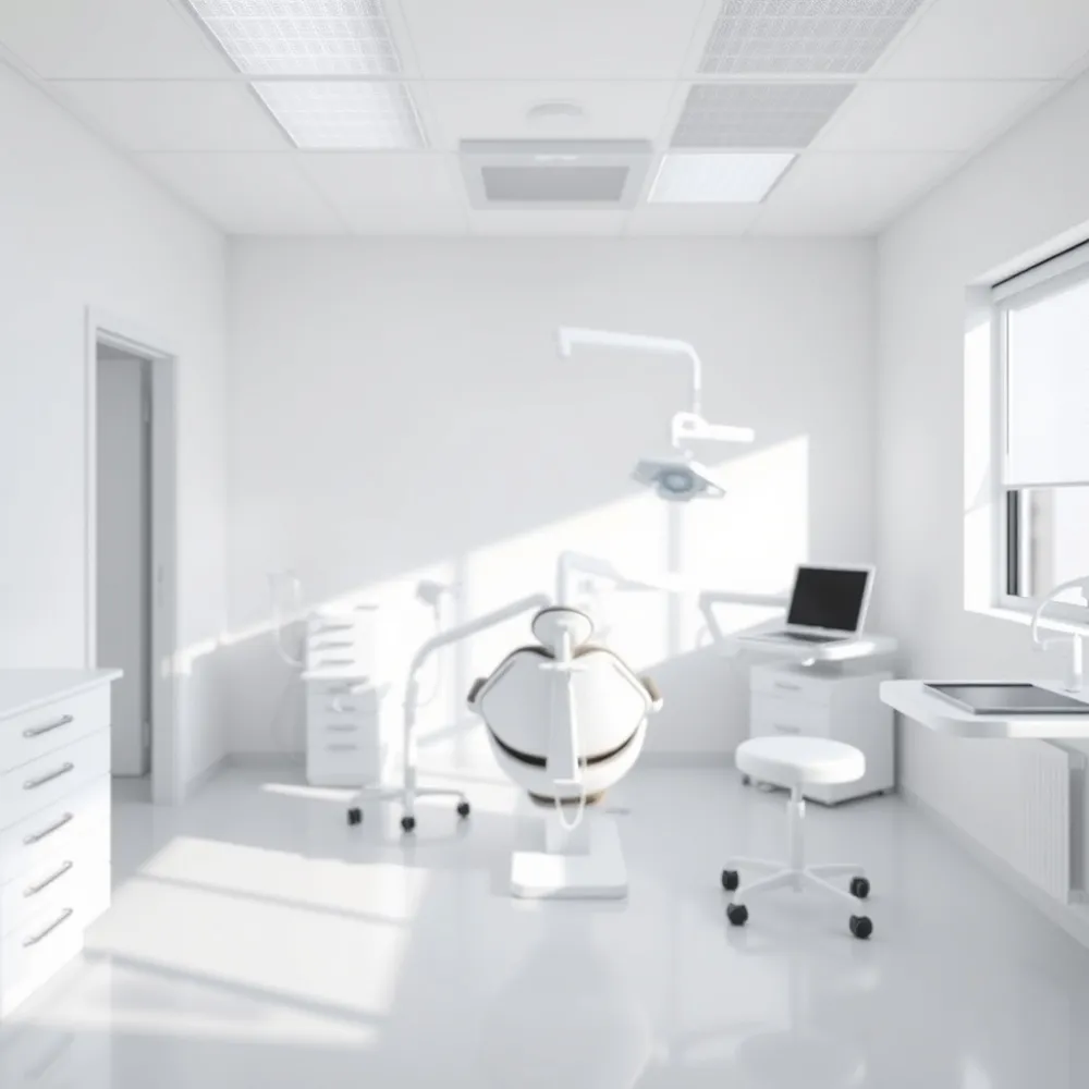
For generations, dental imaging relied on traditional film, a process that required chemical development and time-consuming darkroom procedures. Today, digital radiography has transformed this standard, utilizing sophisticated sensors to capture high-definition images instantly. At drparrella.com, we choose to prioritize these advancements because they allow us to see oral health details with precision while significantly reducing radiation exposure by up to 90 percent compared to older film methods.
We believe that technology is only as valuable as the peace of mind it provides for our local families. Unlike practices that still rely on traditional film-based processing, drparrella.com uses digital systems that capture images with incredible clarity in seconds. This speed means less time in the exam chair and more time focused on your care. Because these images can be magnified and adjusted for contrast, we can identify potential issues, such as small cavities or bone loss, before they develop into serious concerns.
Open, transparent communication is always at the heart of our practice. By displaying your digital dental X-rays on a monitor during your visit, we can walk you through the imagery and explain your treatment plan in real-time. This collaborative approach fosters trust, ensuring you feel fully informed and comfortable at every step. Through the use of modern technology, we are dedicated to providing safer, more efficient care for your entire family.
The Primary Benefits of Digital Dental X-Rays
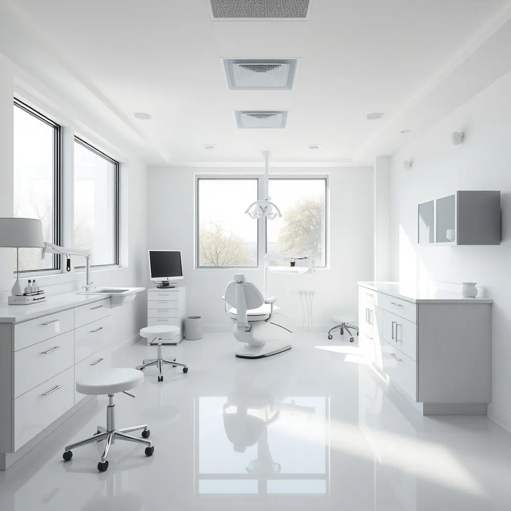
![]() At drparrella.com, we prioritize the health and comfort of our community by utilizing digital radiography to minimize patient exposure. Unlike traditional film-based methods that rely on time-consuming chemical development, digital sensors provide a substantial reduction in radiation exposure of up to 90%. This leap in safety allows for more frequent monitoring when necessary, particularly for children or those tracking specific dental developments.
At drparrella.com, we prioritize the health and comfort of our community by utilizing digital radiography to minimize patient exposure. Unlike traditional film-based methods that rely on time-consuming chemical development, digital sensors provide a substantial reduction in radiation exposure of up to 90%. This leap in safety allows for more frequent monitoring when necessary, particularly for children or those tracking specific dental developments.
Beyond safety, digital technology transforms the consultation experience through immediate image acquisition. Because our digital sensors transmit data directly to a computer monitor, we can review diagnostic data in real-time, removing the waiting periods associated with older darkroom procedures as noted by the NIH. This efficiency supports a collaborative environment where we can walk you through your health data instantly.
The diagnostic clarity provided by digital systems is a major factor in our ability to deliver precise care. Our team uses specialized software to adjust brightness, contrast, and magnification, ensuring we can pinpoint early signs of decay or localized bone loss that might otherwise be missed. By catching these issues early, we can often suggest preventive measures that save you from more invasive procedures later.
- Up to 90% reduction in ionizing radiation compared to traditional film processes.
- Real-time image viewing for immediate patient-dentist consultation.
- Precision adjustment of image contrast and zoom to enhance diagnostic accuracy.
- Environmentally sustainable workflow that eliminates the need for toxic chemical developers.
- Easier digital record management for coordinated long-term dental care.
Understanding Digital Radiography Challenges and Considerations
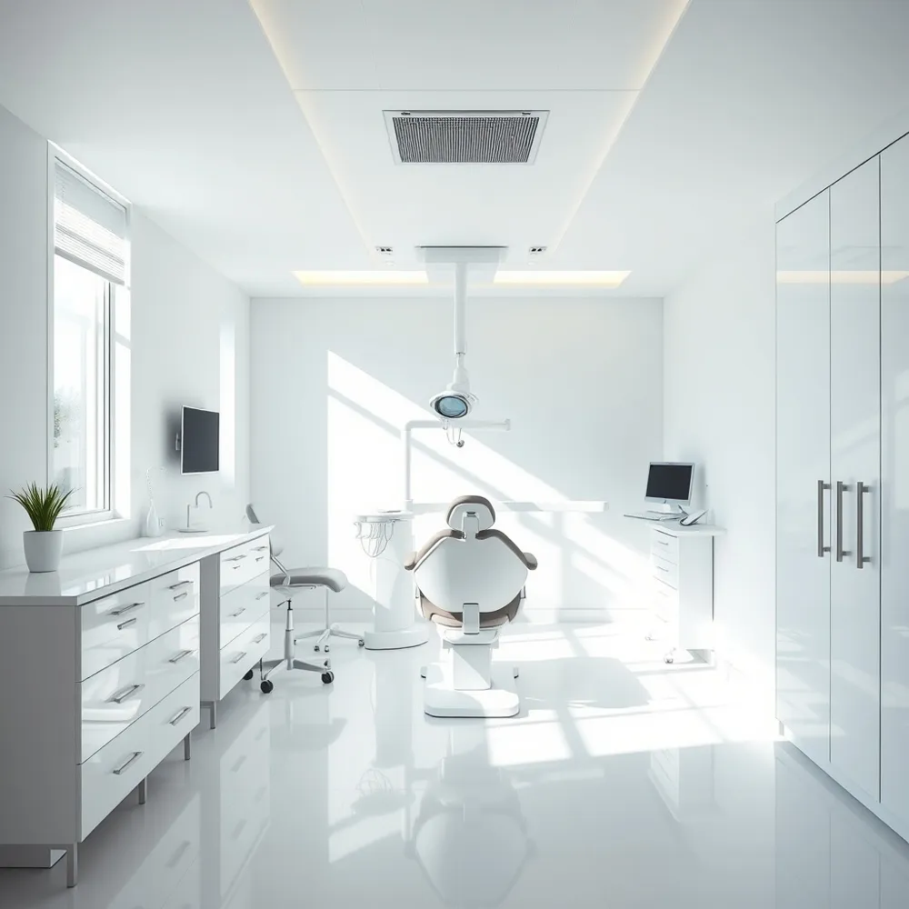
![]() Are there any disadvantages associated with digital radiography in dentistry? While digital radiography is highly effective, some patients may find the digital sensor to be thicker or bulkier than traditional film, which can occasionally cause discomfort or trigger a gag reflex. At drparrella.com, we address these physical anatomy concerns by utilizing smaller, thoughtfully designed imaging sensors that enhance patient comfort during the capture process compared to the bulkier legacy sensors used elsewhere.
Are there any disadvantages associated with digital radiography in dentistry? While digital radiography is highly effective, some patients may find the digital sensor to be thicker or bulkier than traditional film, which can occasionally cause discomfort or trigger a gag reflex. At drparrella.com, we address these physical anatomy concerns by utilizing smaller, thoughtfully designed imaging sensors that enhance patient comfort during the capture process compared to the bulkier legacy sensors used elsewhere.
Additionally, because sensors may have different dimensions than standard film, the field of view per image can sometimes vary during the capture process. Managing these specific imaging constraints requires high-level training to ensure full anatomical coverage without requiring extra exposures. Our team balances these technical variables with precise, patient-focused technique to maintain the high standards of safety established in modern dental imaging practice.
From a practice standpoint, it is important to note that the initial investment for high-quality digital imaging equipment can be significant for a dental office. While some practices might delay this shift, drparrella.com embraces these advanced frameworks to provide faster, safer diagnostic feedback for our local Somerville families. Furthermore, digital systems require robust data management and security protocols to ensure that high-resolution patient images remain protected and easily accessible within the electronic record. We always prioritize your comfort and diagnostic precision, carefully weighing these factors to provide the best possible care for your family's smiles.
How Digital Impressions Enhance Treatment Accuracy
![]() Traditional dentistry often required patients to bite down on trays filled with thick, cumbersome putty to capture the shape of their teeth. At drparrella.com, we have moved beyond these older methods by utilizing advanced intraoral scanners. This technology replaces messy materials with a compact, high-precision camera that creates detailed 3D models of your teeth in minutes. By eliminating the physical strain of trays, we significantly improve your comfort while ensuring we get a perfect capture on the first attempt.
Traditional dentistry often required patients to bite down on trays filled with thick, cumbersome putty to capture the shape of their teeth. At drparrella.com, we have moved beyond these older methods by utilizing advanced intraoral scanners. This technology replaces messy materials with a compact, high-precision camera that creates detailed 3D models of your teeth in minutes. By eliminating the physical strain of trays, we significantly improve your comfort while ensuring we get a perfect capture on the first attempt.
How do digital dental impressions improve the patient experience compared to traditional methods?
Digital dental impressions significantly improve your experience by replacing messy, uncomfortable impression materials with a quick, non-invasive intraoral scanner. This precision technology eliminates the gag-inducing putty of traditional methods while capturing highly accurate 3D images, which results in better-fitting, more comfortable dental restorations. The process is remarkably efficient, allowing for faster chairside visits and an expedited workflow when sending information to our dental laboratory. Furthermore, these digital models allow us to display your oral anatomy on a screen, fostering better communication so you can clearly understand your personalized treatment plan. Ultimately, digital impressions provide a cleaner, more modern, and stress-free way to achieve your dental treatment goals.
Precision is the foundation of long-term oral health, particularly when creating crowns, bridges, and customized implant restorations. Unlike conventional methods that can involve minor distortions during material setting, digital scanning ensures that every restoration matches your unique anatomy with extreme accuracy. Once we reach this level of precision, we can use our digital workflow to send files instantly to our dental laboratories. This speed reduces your waiting time and helps us deliver your final appliance with a much tighter fit and superior aesthetic quality than older restoration techniques typically allowed.
Prioritizing Safety with Established Radiographic Procedures
At drparrella.com, we prioritize your safety by adhering to the ALARA principle, which stands for as low as reasonably achievable. This clinical standard ensures that every diagnostic image we capture is strictly necessary for your care, effectively minimizing your overall radiation exposure.
What standard protective measures are used during dental radiographic procedures?
Our practice utilizes advanced digital radiography, which typically reduces radiation doses by 80% to 90% compared to outdated film-based techniques. While some offices still rely on protective lead aprons and thyroid collars to provide psychological comfort, modern research indicates that these physical barriers are often unnecessary, as they cannot block the internal radiation scatter present during imaging. In fact, relying on outdated shielding can sometimes interfere with image clarity, potentially leading to repeat scans and increased exposure.
It is important to view these procedures in context: the radiation from a standard set of dental X-rays is extremely low, falling well within the levels encountered through natural background radiation—such as from the Earth, granite building materials, or even high-altitude travel per Cleveland Clinic. By following the current recommendations of the American Academy of Oral and Maxillofacial Radiology, our team ensures that your diagnostic process is not only precise but also grounded in the most current safety science.
Inclusive Care and Communication for Every Patient
Providing predictable and compassionate care is a cornerstone of our dental practice at drparrella.com. For patients with sensory needs, including those experiencing visual impairments, fostering a transparent environment is critical to managing anxiety and building lasting trust. We believe that clear verbal communication and respecting personal autonomy are vital to ensuring everyone feels welcome during their treatment.
How should a dental assistant approach a patient with a visual impairment during X-ray procedures?
To support a patient with a visual impairment during X-ray procedures, the dental assistant should first establish clear verbal communication, announcing their presence and describing every step of the process in detail before it begins. It is essential to explain the position of the X-ray equipment and the physical movements required, while avoiding any sudden movements or surprise touches that could cause anxiety. If necessary, you may use tactile guidance by letting the patient touch the non-sharp parts of the equipment to better understand the procedure. Maintaining a calm, reassuring tone throughout the appointment helps build trust and ensures the patient feels secure and informed. Always ask the patient specifically how they prefer to be assisted, as their personal comfort and autonomy are the most important factors in providing compassionate care.
A Commitment to Your Long-Term Oral Health
At drparrella.com, we view advanced technology as more than just an upgrade to our tools. It is a vital component of our mission to provide the Somerville community with transparent, safe, and highly accurate dental care. By moving away from the chemical processing required by traditional film, digital radiography allows us to focus entirely on your comfort and long-term health.
Our practice maintains these high standards because we believe that preventative diagnostics should be as efficient as they are effective. Whether you are due for a routine check-up or need to discuss specific oral health concerns, our team is here to guide you through your options. We invite you to contact our office to schedule your next visit and experience a personalized approach to dentistry that puts your family's well-being first.