
Seeing Beyond the Surface
A visual dental exam can show a great deal, but some areas are difficult to see directly. The spaces between teeth, tooth roots, and supporting bone may hold clues that are hidden from view.
Dental X-rays create images that help a dentist assess these structures and look for concerns such as decay between teeth or changes around a root. They add information to an examination rather than replace it. Dental X-rays can reveal areas that an ordinary visual check cannot fully assess.
Digital images appear on a computer, where their display can be adjusted to help the dentist examine an area of concern. These adjustments do not make the image self-explanatory; a dental professional interprets it alongside your symptoms, history, and examination findings.
At drparrella.com, care is shaped around each patient and their dental needs. Imaging is most useful when it can help answer a clinical question or guide a treatment decision, so the dentist can explain why an image may be recommended and how it could inform your care.
What Dental Images Can Reveal
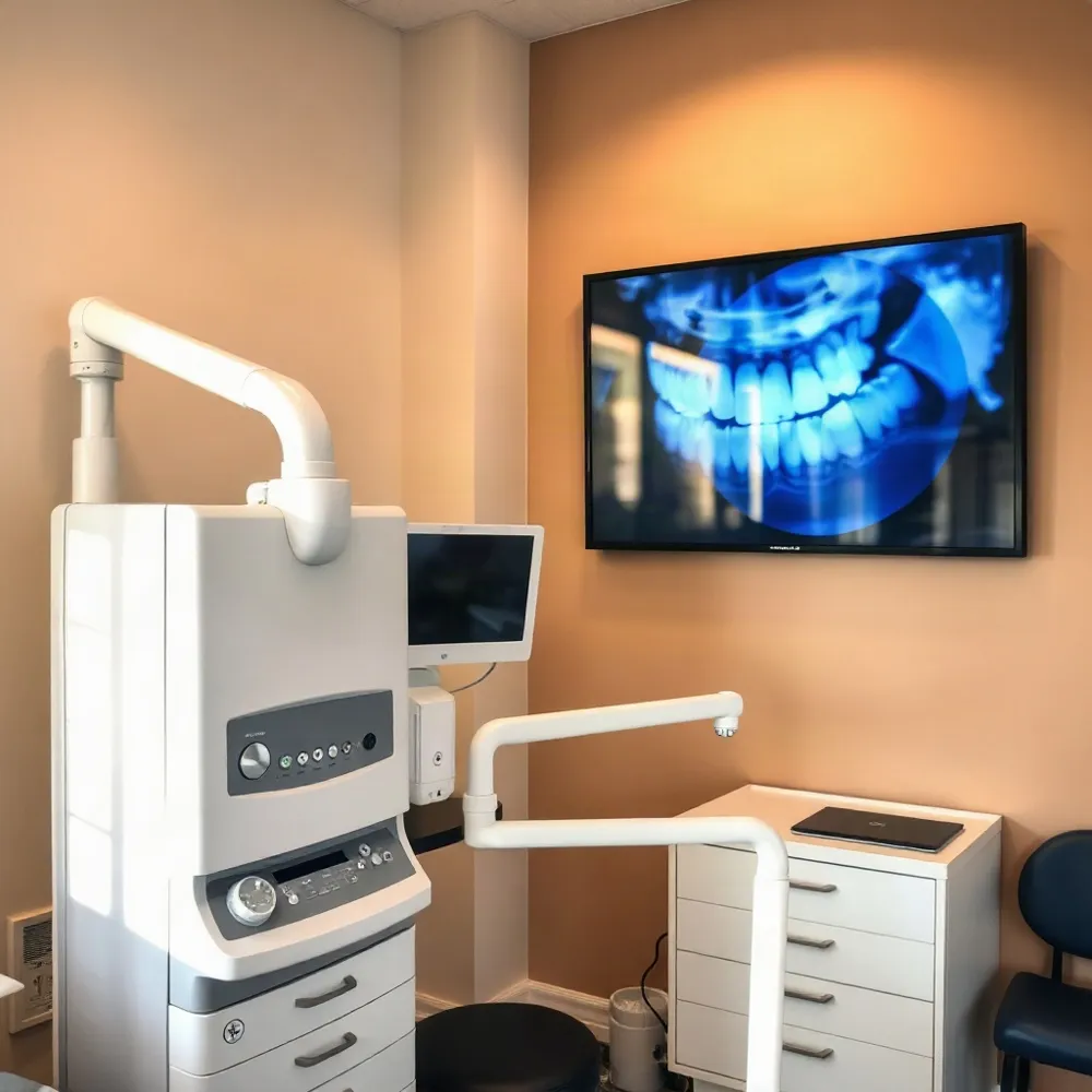 Dental X-rays can show parts of the teeth and jaws that a visual exam cannot reach, helping your dentist investigate concerns and plan care. At drparrella.com, imaging is considered as part of personalized, comprehensive dental care, alongside your exam and health history.
An X-ray image forms because tissues absorb different amounts of radiation. Dense mineralized structures block more X-rays and appear lighter, while less dense areas allow more through and appear darker. This contrast is what makes internal structures visible on a radiograph, as described in this overview of how radiography works.
Depending on the image, a dentist may look for cavities between teeth, decay beneath an existing filling, changes in the supporting bone, or abnormalities near a root that may suggest infection. Different views answer different questions: bitewing images focus on the crowns and spaces between teeth, while periapical images include the roots and nearby bone. Dental X-rays can also help assess unerupted or impacted teeth and support planning for treatments such as implants or orthodontic care.
A dark area is not a diagnosis by itself. Its location and appearance matter, and the dentist interprets it in the context of the clinical examination and your dental history. An image may reveal information that is difficult to see directly, but it cannot guarantee that every problem will be detected or determine treatment on its own.
Images may also help a dentist monitor healing after a procedure. At drparrella.com, recommendations are part of an individualized discussion, so you can understand what an image is meant to clarify and how its findings may affect your care.
Choosing the Right Image
Dental X-rays are not interchangeable versions of the same test. Each type shows a different area, so the choice depends on what the dentist needs to assess and how the result could inform care. The Cleveland Clinic’s guide to dental X-rays describes several commonly used options.
Bitewing images show the crowns of upper and lower teeth in one region of the mouth. They are commonly used to check for decay between teeth and can show nearby bone levels. Periapical images show an entire tooth, from its crown to beyond the root tip, along with the supporting bone around it. These views can help investigate a concern involving a particular tooth or its roots.
A panoramic image provides a broader view of the teeth and jaws. It may help assess impacted wisdom teeth or other jaw concerns, but it generally offers less detail about an individual tooth than an image taken inside the mouth. Cone beam CT is a separate option that produces three-dimensional views. It is chosen for specific clinical purposes, not as a routine replacement for other dental images.
At drparrella.com, care is personalized for local families, and image selection should fit the area of concern and the clinical question. Dr. Anthony P. Parrella’s practice provides comprehensive dental care, giving patients an opportunity to discuss why a particular view is being recommended and how it may guide their care.
A Schedule Based on Your Needs
How often should I have dental X-rays? There is no single schedule that suits every patient. At drparrella.com, care is personalized to each patient, so imaging decisions can take account of your health and examination rather than follow an automatic calendar.
Your dentist may consider current oral health, examination findings, age and dental development, past cavities or gum disease, symptoms, and your risk of decay. The timing and usefulness of earlier images matter too. A recent image from another provider may already answer the clinical question, so it is worth asking whether it can be reviewed before taking a new one.
Some people with healthy teeth, gums, and low decay risk may need images less often. A new symptom, a history of frequent cavities, or another finding during an exam could make an image appropriate sooner. The Cleveland Clinic explains that dental X-ray timing depends on a person's oral health and circumstances, rather than a fixed interval (guidance on dental X-rays).
Routine annual or semiannual X-rays are not automatically necessary for everyone. Before imaging, ask what concern the image is meant to investigate, how the result could affect your care, and whether waiting or using an existing image is reasonable. These questions help make sure each X-ray has a clear purpose and supports your individual care plan.
Radiation and Reasonable Precautions
 Are dental X-rays safe, and can they cause cancer? Dental X-rays use ionizing radiation. At high doses, radiation can affect living cells, so exposure should be managed rather than treated as harmless. In theory, radiation can contribute to cancer risk, but the exposure from routine dental imaging is low, and the added risk is generally considered very small. That does not mean the risk is zero or that every patient needs an image.
Digital dental X-rays use sensors instead of film. Cleveland Clinic reports that they use 80% to 90% less radiation than traditional film X-rays, though actual exposure depends on the image and technique. The ALARA principle means keeping exposure as low as reasonably achievable and taking only images that serve a clinical purpose. A dentist can limit unnecessary exposure by considering your exam and history, choosing the appropriate image, and using proper technique and beam positioning. You can ask what an image is intended to show and whether an earlier image may answer the same question. Cleveland Clinic's overview of dental X-rays explains their uses and general safety considerations.
If you are pregnant or trying to become pregnant, tell your dental team before imaging. Mayo Clinic says dental X-rays typically do not direct the X-ray beam at reproductive organs, and advises discussing your circumstances with a healthcare professional. Mayo Clinic's guidance on X-rays during pregnancy provides more context.
At drparrella.com, care is described as personalized and comprehensive at a longstanding, family-run Somerville practice. A conversation about your health history and the reason for an image can help make the decision fit your circumstances. For more on managing exposure, see radiation and reasonable precautions.
Making an Informed Imaging Decision
 Can I decline dental X-rays, and what should I discuss with my dentist first? You can raise concerns or decline a recommended image. The choice should be a conversation, not a decision made under pressure. Without an image, though, some areas that are difficult to see during an examination may remain unassessed. Dental X-rays can show tooth roots, jawbone, and other structures that a visual exam cannot fully assess, according to the Cleveland Clinic.
Ask what the image is intended to check and how its findings could affect your care. You can also ask whether a recent image might answer the same question, what alternatives are available, and what could happen if imaging is delayed. These questions help connect the recommendation to your circumstances rather than treating X-rays as automatic.
Share any concerns about radiation, pregnancy, health conditions, or ongoing treatment with your dental team. Mayo Clinic advises telling a healthcare professional if you are pregnant or might be pregnant before an X-ray, so the team can consider your circumstances and whether imaging can be postponed or adjusted. Mayo Clinic’s guidance on X-rays during pregnancy offers general information, while your dentist can discuss your specific situation.
At drparrella.com, care is described as personalized and comprehensive, with an emphasis on clear communication. That means patients should have room to ask questions and understand the reasons for a recommendation. Image findings are considered alongside your exam and health history, not in isolation. Ask your dentist to explain the results in everyday language and how they relate to possible next steps. You can also review how the practice approaches informed choices before treatment.
Understanding the Cost
How much do dental X-rays cost? Without insurance, they may cost roughly $25 to $250, though the fee depends on the image type, how many images are taken, and the dental office. A single image may cost less than a full set or a panoramic X-ray.
Dental insurance may cover some or all of the cost, depending on your plan’s coverage limits and how often it pays for X-rays. Before imaging, ask the practice and your insurer for an estimate. If the cost is a concern, tell the dental team so you can discuss options.
At drparrella.com, care is provided by a family-run Somerville practice focused on personalized dental care. The reason for each image should be clear: recommended X-rays should help assess your oral health or guide treatment, rather than add an unexplained expense. For more on the factors behind the estimate, see dental X-ray costs and access.
Images That Support Personal Care
Dental X-rays can show areas that are difficult or impossible to examine directly, but their value lies in answering a specific clinical question. An image is one part of an assessment, considered alongside your symptoms, dental history, and examination.
When an image is recommended, ask what it is intended to show, what the findings mean, and how they may affect your care plan. At drparrella.com, personalized, comprehensive care and clear communication help local families discuss recommendations with their dental team and make informed decisions.