A Modern Look Inside Your Smile
Digital X-rays represent a major advancement in dental care, allowing dentists to see hidden issues like early cavities, bone loss, and infections that are invisible during a standard exam.
Practices like drparrella.com use this technology because it offers exceptional safety. Digital systems expose patients to 80% to 90% less radiation than traditional film-based methods, making routine imaging both effective and remarkably low-risk.
Understanding the Radiation Reduction
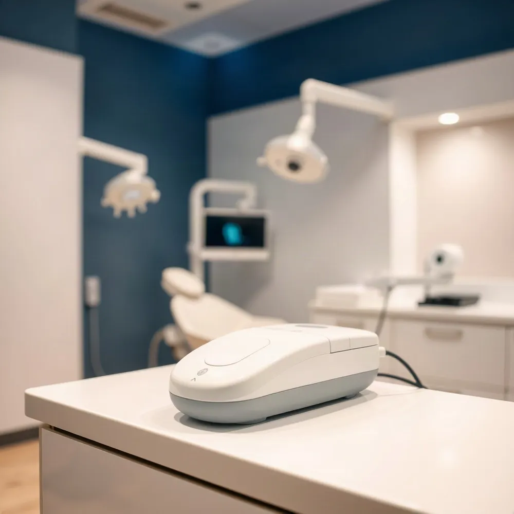 One of the most common questions patients ask is whether digital X-rays use less radiation than traditional film-based ones. The answer is clear: digital sensors require up to 80-90% less radiation than conventional film to produce a diagnostic-quality image. This dramatic reduction is possible because digital sensors are far more sensitive to X-ray photons, capturing a usable image with a much shorter exposure time.
To put this in concrete terms, a set of four digital bitewing X-rays exposes you to about 0.005 millisieverts (mSv) of radiation. That is roughly the same amount you absorb from a single day of natural background radiation from the sun, the earth, and everyday building materials. It is about twenty times less than a standard chest X-ray (0.1 mSv) and roughly 2,000 times less than a CT scan of the abdomen.
Because the exposure is so minimal, the American Dental Association updated its guidance in 2024, stating that lead aprons and thyroid collars are no longer necessary for routine digital dental X-rays. At drparrella.com, we follow these modern safety guidelines, using digital sensors that allow us to capture the images we need with the lowest possible dose, giving you both a thorough exam and peace of mind.
How Many X-Rays Are Safe in One Visit?
A full diagnostic set of X-rays, such as a full mouth series of 14 to 20 images, can be taken safely in a single visit. Modern digital sensors require 80% to 90% less radiation than traditional film, so even a complete series delivers a very low cumulative dose. At Dr. Parrella's practice, digital X-rays make it possible to gather all the diagnostic information your dentist needs without exceeding minimal exposure levels.
There is no strict daily limit for necessary diagnostic purposes. Dentists follow the ALARA (As Low As Reasonably Achievable) principle, which guides them to prescribe only the X-rays needed for an accurate diagnosis and to use techniques that further reduce the dose. A set of four bitewing digital X-rays exposes a patient to about 0.005 millisieverts, roughly the same amount of radiation absorbed from a normal day in the sun.
How Often Should You Schedule Dental X-Rays?
The frequency of dental X-rays depends on your individual health needs. For most patients with good oral health, bitewing X-rays are recommended every 12 to 24 months to check for cavities between teeth. A full mouth series or panoramic X-ray is typically taken every 3 to 5 years to monitor overall bone health and detect issues below the gumline.
Your dentist at Dr. Parrella's office will tailor the schedule to your age, risk factors, and oral health history. Patients with gum disease, frequent cavities, or ongoing treatments may need X-rays more often. This individualized approach ensures you receive the diagnostic benefits without unnecessary exposure.
| X-Ray Type | Typical Frequency | What It Detects |
|---|---|---|
| Bitewing | Every 12-24 months | Cavities between teeth, bone density changes |
| Full Mouth Series | Every 3-5 years | All teeth, roots, and surrounding bone |
| Panoramic | Every 3-5 years | Jaws, sinuses, impacted teeth, overall oral structures |
Key Advantages of Digital Radiography
 Digital radiography uses electronic sensors instead of film, producing images that appear on a computer screen in seconds. This shift delivers tangible benefits for both diagnosis and patient experience.
Superior Image Quality and Immediate Results
Unlike static film, digital images can be enlarged, magnified, and adjusted for contrast and color. This helps detect early-stage cavities, subtle bone loss, and hidden infections that might be missed on traditional film. At drparrella.com, we use this capability to examine every detail thoroughly.
Because the image is available instantly, your dentist can identify problems and discuss treatment options during the same visit. There is no waiting for chemical development, which reduces time in the chair and eliminates retakes due to incorrect exposure.
Storage and Sharing. Digital files are stored electronically in your patient record, making it easy to transfer them to a new dentist or share with a specialist for a second opinion. This eliminates repeat X-rays when you switch providers, keeping your lifetime exposure low.
Eco-Friendly Process. Digital radiography removes the need for darkroom chemicals and lead foil used in film development. This reduces hazardous waste and makes the practice safer for the dental team and the environment.
Essential for Comprehensive Care
High-resolution digital X-rays are essential for planning crowns, root canals, and orthodontic treatment. They reveal the position of tooth roots, nerve canals, and surrounding bone, guiding precise procedures. For children, regular digital images allow monitoring of developing teeth and jaws, helping identify alignment issues before they become complex problems.
These capabilities align with the ALARA principle (As Low As Reasonably Achievable), ensuring that the lowest possible radiation dose is used to obtain the diagnostic information needed.
Types of Dental X-Rays and Their Uses
Dentists use different types of X-rays depending on what they need to see, each designed for a specific diagnostic purpose.
Intraoral X-Rays (Sensor Inside the Mouth)
Bitewing. Captures the crowns of upper and lower teeth in a single view, showing cavities between teeth and changes in bone height caused by gum disease.
Periapical. Shows the entire tooth — from crown to root — along with the surrounding bone structure. These are used to detect abscesses, infections, and bone loss linked to individual teeth.
Occlusal. Provides a broader view of the floor of the mouth or palate, helping locate impacted teeth, jaw fractures, or extra teeth.
Extraoral X-Rays (Sensor Outside the Mouth)
These images capture larger areas and are essential for treatment planning rather than spotting single-cavity issues.
Panoramic. A single sweeping image that shows all teeth, jaws, sinuses, and surrounding structures. It is commonly used to plan extractions, orthodontic work, or dentures.
Cephalometric. A side view of the head used in orthodontic treatment planning to assess jaw relationships and tooth alignment.
Cone Beam CT (CBCT). A 3D image of the teeth, jaws, nerves, and sinuses, often used for precise implant placement or evaluating complex oral anatomy.
The main difference between periapical and panoramic X-rays is scale. A periapical image provides a detailed, close-up view of one or two teeth, making it ideal for diagnosing root infections or abscesses. A panoramic X-ray offers a broad overview of the entire mouth in a single shot, which is useful for planning braces, dentures, or bone grafts. At drparrella.com, the team selects the appropriate type of X-ray for each patient's needs, ensuring the right balance of diagnostic detail and minimal radiation exposure.
Addressing Patient Concerns and Side Effects
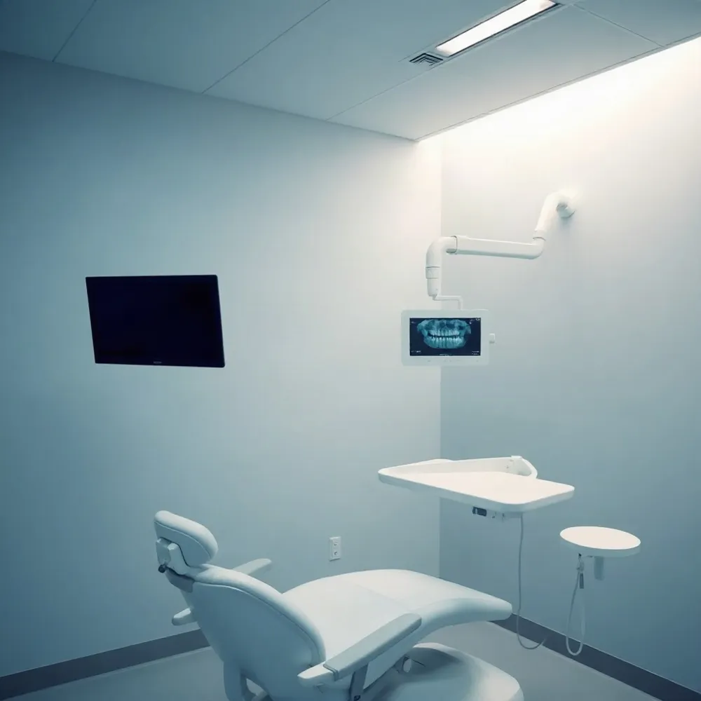 For the vast majority of patients, the risk of side effects from a routine dental X-ray is essentially nonexistent. The radiation exposure from a set of digital dental X-rays is often less than what you receive from natural background radiation in a single day. While ionizing radiation in large doses can be harmful, modern technology minimizes exposure to a negligible level.
Some patients may experience minor and temporary discomfort unrelated to radiation. This can include slight discomfort from the positioning of the X-ray sensor, especially since digital sensors can be thicker than traditional film. A temporary gag reflex during placement is also possible.
Safety for Pregnant Patients
The safety of dental X-rays during pregnancy is well-established. The Cleveland Clinic notes that both the American Dental Association and the American Pregnancy Association agree the risk to the fetus is minimal. Most dentists take extra precautions, but emergency X-rays during pregnancy are not a cause for concern.
Main Disadvantage. The primary disadvantage of digital imaging is the higher initial cost for the necessary equipment, including sensors and supporting software. This cost is absorbed by the practice and does not result in higher fees for patients.
The 15% Rule. A traditional radiography principle states that decreasing the kilovoltage peak (kVp) by 15% requires doubling the milliampere seconds (mAs) to maintain consistent exposure. This guideline is primarily used when adjusting image contrast, but modern digital systems have made it less critical.
The significant diagnostic benefits of dental X-rays far outweigh any minimal risks. At drparrella.com, modern digital systems are used to ensure patient comfort and safety, providing clear images with exceptionally low radiation doses.
Peace of Mind Through Modern Technology
Digital X-rays are a cornerstone of modern, patient-centered dentistry. At drparrella.com, this technology provides a safer, faster, and clearer diagnostic experience, allowing your dentist to detect cavities, bone loss, and other issues early. This commitment to advanced tools and personalized care ensures every visit is both comfortable and thorough.