
What Routine Exams May Miss
A visual dental exam can reveal a great deal, but some areas are difficult to assess by looking alone. Dental images offer another view, showing structures between teeth and beneath the gumline, including roots and supporting bone. They may help identify decay, bone changes, or other concerns that are not yet obvious during an examination, as Cleveland Clinic’s guide to dental X-rays explains.
Images do not replace an exam or automatically mean treatment is needed. At drparrella.com, decisions about imaging are part of personalized care: a dentist considers symptoms, dental and medical history, examination findings, and any planned treatment before recommending a type of image or its timing. Patients can ask what the image is intended to show and how the result might affect care.
Types of Images and Hidden Findings
 Dental X-rays, also called radiographs, give your dentist a view of teeth, roots, and jawbone areas that a regular exam cannot fully show. Different images answer different questions, so the choice depends on your oral health, symptoms, and treatment needs.
Bitewing images help check for decay between teeth and below the gumline. Periapical images show a tooth from crown to root and the surrounding bone, which can help assess concerns near the root. A dental X-ray guide describes these and other common views.
For a broader view, panoramic images capture the teeth and jaws together. Occlusal views show a larger area of the upper or lower jaw, while cephalometric images provide a side view often used in orthodontic planning. Cone beam CT scans create three-dimensional images and may be selected for specific planning needs, such as dental implants.
Images may reveal decay between teeth or beneath fillings, bone loss associated with gum disease, impacted teeth, or signs of infection near a root. They can also help assess changes around existing restorations. At drparrella.com, imaging is part of personalized, comprehensive care, with findings considered alongside the patient’s history and examination.
Finding a problem before it progresses may leave different treatment options available, but an image alone does not determine care. Your dentist interprets it with your symptoms and exam findings, then explains whether treatment or follow-up is appropriate. Images can also help monitor healing after certain procedures.
Digital Imaging and Radiation Safety
 Digital X-rays use electronic sensors rather than film, so images can be reviewed soon after they are taken. A dentist can enlarge or adjust an image for closer inspection, and digital files can be stored or shared with other providers. These features support clear discussion of findings during a visit.
Digital systems may use less radiation than film systems, but the difference depends on the equipment and technique. Both methods use ionizing radiation, and digital imaging is not radiation-free. Dental X-rays involve a small amount of radiation, and the risk from an individual image taken for a clinical reason is considered low. The Cleveland Clinic’s overview of dental X-rays explains that images can help identify issues that a visual exam may not show.
Safety depends on using images when they can inform diagnosis or care, rather than following a fixed schedule for everyone. Dental teams can limit exposure by directing the beam to the area being examined, positioning the sensor carefully, and checking whether usable images from a previous visit are available. Careful positioning can also help avoid unnecessary repeat images.
The American Dental Association’s 2024 recommendations no longer call for lead aprons and thyroid collars as routine measures for dental X-rays. Practices may still use additional precautions, and your dental team can explain its approach. Tell your dentist if you are pregnant or may be pregnant, or if radiation raises concerns for you.
There is no universal safe number of dental X-rays or single interval that suits every patient. Age, dental history, risk of cavities or gum disease, current symptoms, and planned treatment all help determine whether an image is useful. At Dr. Parrella’s family-run Somerville practice, imaging decisions are part of personalized care, and patients can ask what an image is expected to show and whether earlier images can answer the question.
Reading Images Alongside Symptoms
An X-ray can show changes around a tooth root or in nearby bone, but it cannot show the tooth's nerve directly or confirm that the nerve is healthy. These findings may point to a problem inside the tooth, but they are only one part of the picture.
To assess the pulp, the inner tissue containing nerves and blood vessels, a dentist considers symptoms and examination findings alongside the image. Cold or electric pulp tests can help assess how the pulp responds. Dental X-rays may show related changes around roots, but images do not independently confirm every condition.
Tell your dentist if you have lingering pain, unusual sensitivity, swelling, or a tooth that feels different. An evaluation can help identify the cause and determine whether imaging or other tests could guide diagnosis and treatment timing. At drparrella.com, care is provided by a family-run practice in Somerville, where discussion of symptoms and findings can inform decisions tailored to each patient.
For more on what images can and cannot reveal, see how dental images are read alongside symptoms.
Personalized Decisions for Every Patient
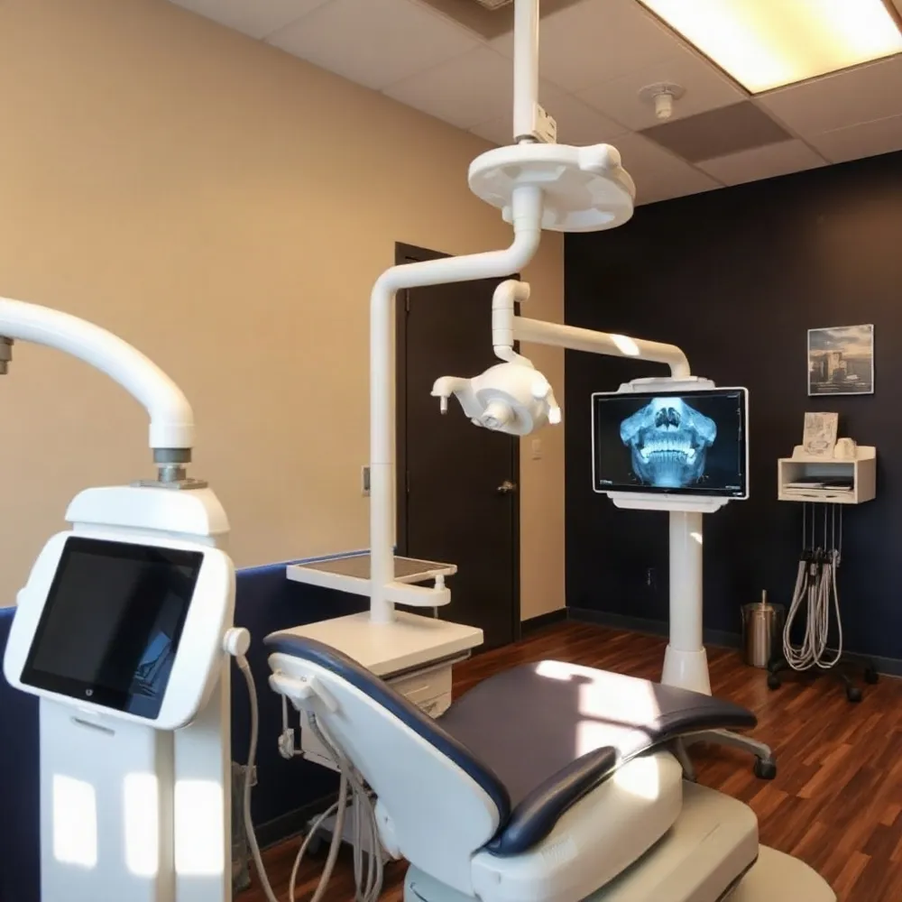 For children, dental images should be recommended for a specific reason, not simply because a routine schedule says it is time. The dentist considers age, dental development, symptoms, past decay, examination findings, and whether the image is likely to change care. Imaging may help track incoming teeth or investigate a concern that cannot be assessed by looking alone. Rutgers School of Dental Medicine describes how radiographs can support assessment of children’s dental development and hidden decay.
Parents can ask what the image is intended to show and whether recent images from another office are available. At drparrella.com, care is described as personalized and comprehensive, making a child’s history and examination findings relevant to the discussion rather than relying on a one-size-fits-all approach.
Patients may decline recommended X-rays. Before deciding, ask why the dentist advises imaging, what could be harder to detect without it, and how waiting might affect treatment planning. Cleveland Clinic’s guidance on dental X-rays explains that images can reveal problems such as decay between teeth, infection, or bone loss that a visual exam may not show.
You can also ask whether another approach or postponement is reasonable. If a treatment cannot be planned safely without the information an image provides, the dentist may recommend limiting or delaying that care. A clear conversation helps patients and families weigh the reason for imaging against their concerns and decide on an appropriate next step.
Understanding the Cost of Dental Images
Dental X-ray costs vary with the type and number of images, your location, and your insurance coverage. As a general guide, bitewing or periapical images may cost about $40 to $120, while a full-mouth series may range from roughly $175 to $430. Panoramic images may cost more than individual images because they show a broader area.
These ranges are estimates, not guaranteed prices. Your out-of-pocket cost may be lower if your plan covers diagnostic X-rays, but benefits and frequency limits differ. Ask the dental team for an estimate based on the images recommended for your needs, and check your insurer’s coverage before the appointment. Recent images may also help the team decide whether new ones are needed. Learn what can affect the cost of dental images.
A Clear Conversation About Care
Digital X-rays are a diagnostic tool, not an automatic part of every visit. Their value depends on the question a dentist needs to answer and each patient’s circumstances. At Dr. Parrella’s family-run Somerville practice, care is personalized, so patients can discuss why an image may be useful before deciding.
Patients and families can ask what an image is expected to show, how its findings could affect care, and whether recent images from another office can be reviewed first. These conversations help the dental team avoid unnecessary duplication and explain the options clearly. Informed decisions begin with understanding the reason for a recommendation.