Why Dental Imaging Matters
A dental exam can reveal a great deal, but it cannot show every surface between teeth or structures beneath the gums. Dental X-rays let a dentist assess areas such as tooth roots and jawbone, and can help identify concerns that are not visible during a routine visual check. They support an examination rather than replace it, and the dentist interprets images alongside your symptoms and oral health history. Cleveland Clinic’s guide to dental X-rays explains what these images can help evaluate.
This section compares how film-based and digital methods capture, display, and store dental images. Film must be developed before it can be viewed; digital systems capture images electronically for review on a computer. Either approach may provide useful information, but the right image and method depend on the clinical question and the patient’s needs. In a family-run practice such as drparrella.com, those choices belong in a clear conversation between patient and dentist, not in a one-size-fits-all schedule.
How Digital and Film Images Differ
Digital dental X-rays capture an image with an electronic sensor or imaging plate and display it on a computer. Traditional film X-rays expose photographic film, which must be developed with chemicals before the image can be viewed. Both methods provide images dentists can use alongside an examination to assess oral health.
With a direct digital sensor, an image is usually available within seconds. Phosphor imaging plates also create digital images, but the plate must first be scanned. Film takes longer because it must be processed. In a family practice such as drparrella.com, quicker access can make it easier to review an image with a patient during the visit.
Digital software can enlarge an image or adjust its brightness and contrast, helping the dentist inspect an area of interest. Electronic files can also be stored, retrieved, and shared with another dental professional. These tools support review and coordination, but they do not guarantee a more accurate diagnosis. Image quality and the clinician’s interpretation still matter. A study comparing scanned dental films with their originals found comparable clarity under its specific conditions, not proof that every digital system performs the same way (study of scanned radiographs and conventional films).
Digital systems may use less radiation than film, though the amount depends on the equipment and imaging protocol. Both approaches use X-rays, so the decision to take an image should reflect the patient’s needs. drparrella.com’s family-centered care is grounded in discussing findings and care choices with patients, rather than treating the format alone as a measure of diagnostic value.
Benefits and Practical Limitations
Digital images are usually ready for review soon after capture. A dentist can enlarge them or adjust brightness and contrast to inspect an area, then store the files in a patient record or send them to another provider for a referral or follow-up. These features make it easier to review findings and discuss them during an appointment. Image adjustments can support that conversation, but they do not replace a dentist’s interpretation of the image alongside an exam and the patient’s history.
Digital receptors may require less radiation than film, but the amount depends on the equipment and imaging protocol. Cleveland Clinic’s guide to dental X-rays discusses digital and film methods; a lower exposure should not be assumed for every image or patient. Digital imaging also avoids the film-processing chemicals used in conventional workflows.
There are practical limits, too. Digital systems need specialized equipment and software, along with maintenance and staff training. A sensor may feel uncomfortable in the mouth, and placement difficulties or image artifacts can affect whether a picture is useful. If an image is unclear, the dentist must decide whether another view is needed. At Dr. Parrella’s family-run practice, imaging decisions fit within personalized, comprehensive care, with findings considered in the context of each patient’s needs.
For more on how on-screen review and electronic image sharing fit into patient care, see the connected, eco-friendly practice.
What are the benefits and limitations of digital dental X-rays?
They offer quick access, adjustable views, and convenient storage and sharing, which can support review, planning, and follow-up. Their limits include equipment and maintenance needs, possible image-quality problems, and sensor-placement discomfort. A dentist interprets each image in context and recommends imaging when appropriate.
Radiation and Individualized Imaging
Dental X-rays use ionizing radiation whether the image is captured on film or with a digital sensor. Digital systems may require less exposure, but the amount varies with the equipment, settings, image type, and patient. Cleveland Clinic’s guide to dental X-rays describes digital imaging as using less radiation than traditional film, but that comparison does not mean every digital examination has the same dose.
There is no single imaging schedule or number of X-rays that fits everyone. A dentist considers your oral health, symptoms, dental history, and risk of developing disease when deciding whether images could help. The aim is to use the lowest exposure that can produce clinically useful images and to avoid X-rays that are not indicated.
At drparrella.com, imaging decisions are part of individualized dental care, rather than a fixed calendar applied to every patient. If an image is recommended, you can ask what the dentist wants to assess and how the result may affect your care.
How safe are dental X-rays, and how often might I need them?
Dental X-rays use a relatively low amount of radiation, though exposure varies and no universal monthly limit or fixed schedule applies to every patient. Your dentist can explain the reason for an image and discuss concerns before it is taken. Share personal circumstances, including pregnancy or other health concerns, so the recommendation can be considered in context. If you have previous images, mention them as well, since they may help the dental team assess what is needed.
What Happens During Digital Imaging
 The process depends on the area the dentist needs to examine. For an image of a few teeth, a dental professional places a small sensor inside the mouth. For a broader view, the equipment may sit outside the mouth while it captures the image. Dental X-rays can show areas that a visual exam cannot reach, including tooth roots and the jawbone.
How are digital dental X-rays taken?
The dental professional positions the sensor or helps you stand or sit in the right place. You may be asked to stay still or gently bite on a holder to keep the sensor steady. The X-ray unit briefly exposes the area, and the sensor records the image. A computer then displays it for review, usually shortly afterward.
Sensor placement can feel awkward, and comfort varies with the equipment and the person. Tell the dental team if you have a sensitive gag reflex or find it difficult to hold the sensor in place. At drparrella.com, imaging is part of the practice’s personalized dental care, so patients can raise questions about positioning or the reason for an image during their visit.
The dentist chooses the number and type of images according to the clinical question and your oral health needs, rather than taking a fixed set for everyone. Images can then be reviewed with you to explain what they show and what, if anything, may need follow-up. A walk-through of digital imaging offers more detail about the general process.
Choosing a View for the Question
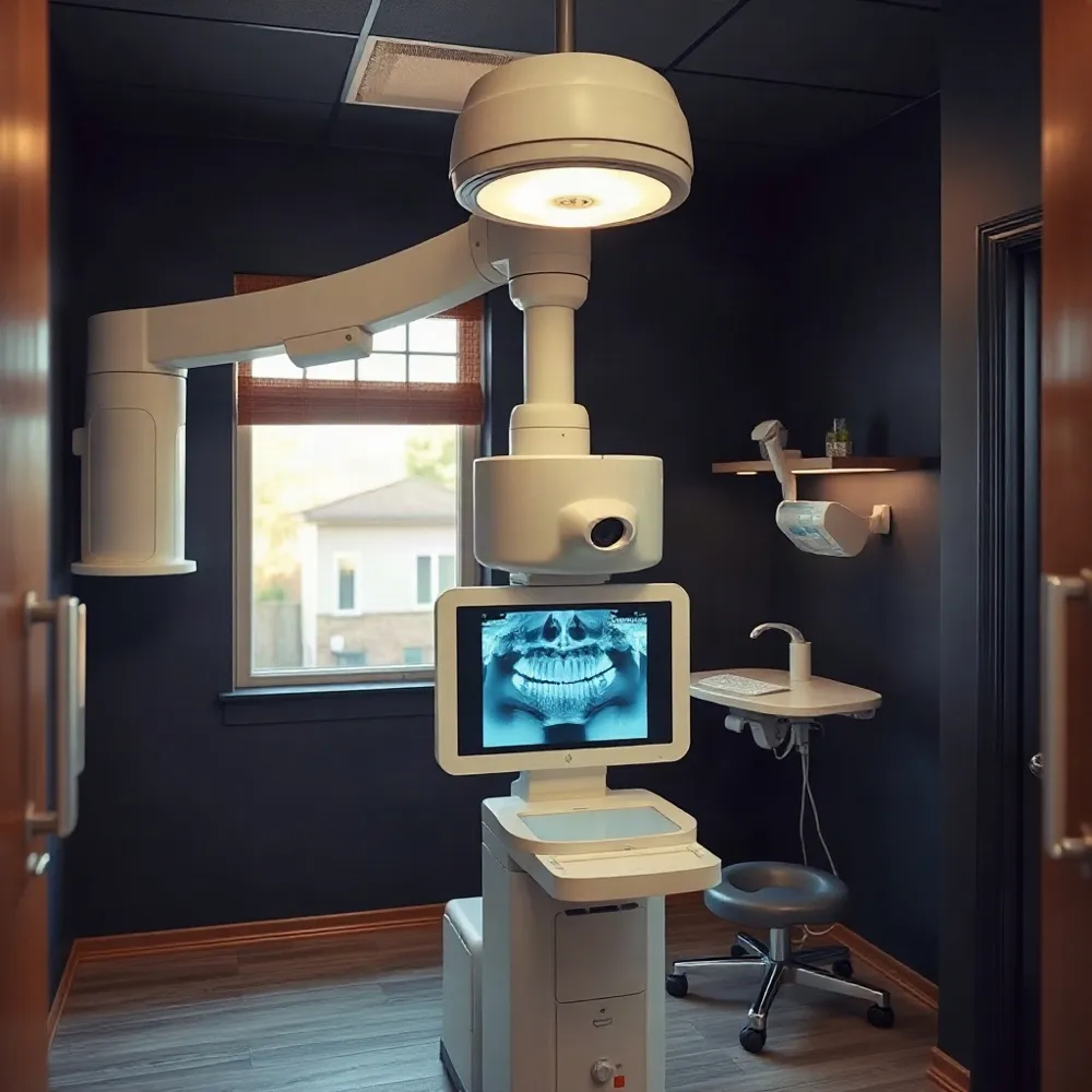 Dental X-rays come in different views because each is designed to answer a different clinical question. A periapical image focuses on one or a few teeth, while a panoramic image shows a wider area of the mouth and jaws.
What is the difference between a periapical and a panoramic dental X-ray?
A periapical X-ray shows a tooth from its crown to the root tip, along with the nearby bone. It can help a dentist assess a concern involving a particular tooth or root, such as a possible infection or a change around the root.
A panoramic X-ray provides a broad, two-dimensional view of the upper and lower teeth and jaws. It can help assess overall dental development or the position of impacted teeth. The Cleveland Clinic’s guide to dental X-rays describes how dentists use different images to examine teeth and jaw structures and plan care.
A wider view does not show the same fine detail as a focused periapical image. The dentist chooses the image that best fits the area or concern being assessed, rather than assuming one type is right for every visit. Patients at drparrella.com can ask what the selected view is intended to show and how it relates to their exam.
For a closer look at how other dental views serve different purposes, see this guide to periapical and panoramic imaging. Understanding the reason for an image can make it easier to follow the dentist’s findings and discuss possible next steps.
Questions, Choices, and Shared Decisions
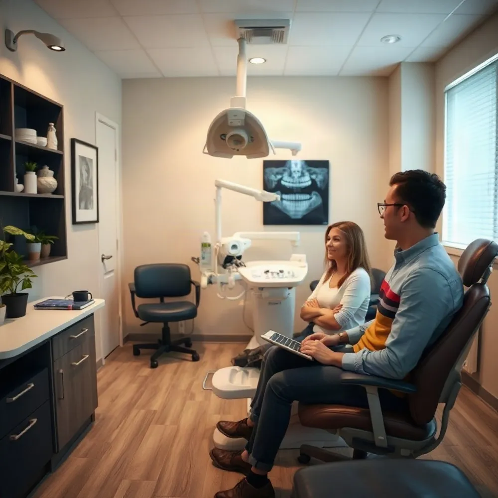 Yes, you can decline dental X-rays. Your dentist should explain why images are recommended, answer your questions, and discuss what care is possible if you choose not to have them. At drparrella.com, patient-centered care means making room for clear conversations about your concerns and choices.
An examination can show only what is visible in the mouth. X-rays may reveal decay between teeth, infection around a root, bone loss, or impacted teeth that a visual check can miss. Without images, some concerns may go undetected, and decisions about treatment may be less certain. Dental X-rays help dentists assess areas beyond the reach of a routine visual exam.
If radiation, pregnancy, cost, or timing is on your mind, say so before imaging. Ask whether a different time or another approach would suit your circumstances. The dentist can explain the expected benefits and risks in your situation, then discuss which care options remain available. A shared decision should not involve pressure or a promise that an examination can proceed unchanged without the recommended images.
You may also find it helpful to review patient safety and precautions before your appointment. Bring your questions, and let your dentist know about relevant health changes so you can decide together how to proceed.
A Conversation About Your Care
Digital and film X-rays differ in how they capture, display, and store images, but neither format alone determines which view is right for you. Dental X-rays can help assess areas that a visual exam cannot show, and the choice depends on your oral health and the question your dentist needs to answer. Cleveland Clinic’s guide to dental X-rays explains their uses and the need to consider individual circumstances.
If an image is recommended, ask what it is intended to assess and how the result could affect your care. You can also ask whether an existing image may be useful, whether another approach is suitable, and when imaging should take place. At drparrella.com, personalized care means discussing your circumstances and making room for questions before deciding together on next steps.